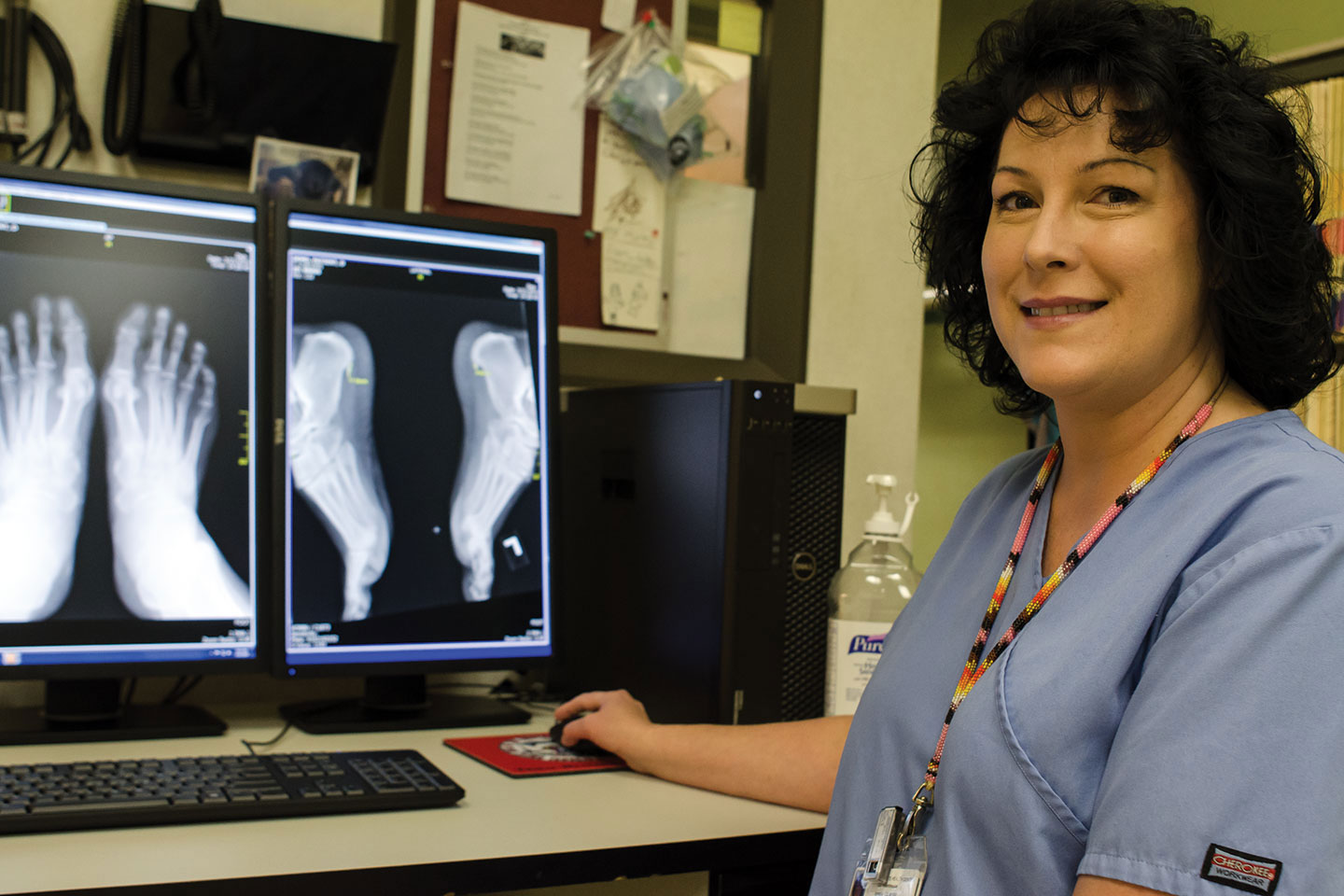
Digital X-ray technology has come to the Southern Ute Health Center, making available next-day readings that previously took three or four weeks to complete.
The new process does away with processing and reading of film images in the same way consumer cameras did away with rolls of film a decade ago, said Dr. Michael Torres, interim chief medical officer. Now, the new equipment takes digital images that are then made available via software for radiologists to read instantly, he said.
“This is a tremendous improvement,” Torres said. “We’re getting readings much faster than before.”
Under the previous arrangement, a local radiologist would pick up films from the clinic and typically return readings several weeks after the images were made. If the clinic needed an immediate reading, an employee would drive the film to Mercy Regional Medical Center and the tribe would pay a premium for the service, Torres said.
The new process not only works much faster – it’s also less expensive, Torres said. The tribe has signed a contract with Tulsa, Okla.-based Diagnostic Imaging Associates Inc., which employs more than 30 board-certified radiologists, to do the readings overnight at a rate less than what it had paid before. If the clinic needs a reading sooner, a staffer can also send an image directly to DIA and request an immediate response.
Health Center patients can expect the same experience as before when being imaged, Torres said.
“The patient doesn’t know the difference; we do,” he said.
As an added benefit, he said, those reading the images can manipulate them in ways impossible using film. Imagine all the changes you can make to your personal digital photos that you couldn’t make with a film camera: Those are the things your doctor can now do with your medical images, Torres said.
“You can get rid of artifacts, increase or decrease the contrast, increase or decrease the brightness,” he said, adding that such simple tweaks can help doctors determine, for instance, the depth of a problem spot within the body.
Other benefits to the new way of doing things: certain imaging procedures use just half of the radiation they previously did, the Health Center no longer has to store hazardous chemicals for developing film and cleaning the equipment, and the images themselves are secured on tribal servers and only shared on a limited basis with authorized personnel.